Histology
Fixation
Tissue should be placed in the fixative of choice as soon as it is removed from the body/animal or as soon after death as practicable. There are a variety of fixatives available with no one fixative regarded as perfect. The most commonly used fixative is formaldehyde which is supplied as a solution of approximately 37% formaldehyde gas in water. This concentration is regarded by histotechnologists as 100% formalin when preparing fixative solutions. The usual working solution is 10% neutral buffered formalin, the formalin being buffered with phosphate salts. The buffering prevents the formation of formic acid which produces formol-haeme pigment in the tissues after prolonged periods. This is an artefact which needs to be removed prior to staining. Formalin fixes by cross-linkages formed in the proteins. These cross-linkages do not unduly affect the structure of the proteins so that the antigenic sites are still available for immunohistochemistry.
Formalin penetrates well but fixes slowly - approximately one mm per hour. It causes little or no shrinkage in the tissue and any routine staining method can be used after formalin fixation. Formalin has an unpleasant smell and needs to be handled with care. It is a strong eye, skin and mucous membrane irritant, is a known skin sensitiser and is an animal carcinogen. Another common fixative in use is Bouin's fluid which is a mixture of saturated aqueous picric acid, formaldehyde and glacial acetic acid. It has an unknown mechanism of action, penetrates well and fixes rapidly. However, tissue left in Bouins for prolonged periods will suffer excessive shrinkage. Generally, 3-6 hours is adequate, depending on tissue size.
Tissue should then be transferred to 70% alcohol (ethanol) which helps leach out the picric acid. Picric acid left in the tissue block can have a degenerative effect after long term storage. Several changes of alcohol over 4-6 hours are needed. After removal of the picric acid, staining results are excellent. (The tissue will always remain yellow). Care must be taken when preparing Bouin’s fluid as the picric acid powder must not be allowed to dry out - it is explosive when dry. Always ensure that there is a good volume of distilled water in the bottle before returning to the chemical store. Another factor to be aware of is that everything picric acid touches turns yellow. This includes clothes, from which it is almost impossible to remove, floors, shoes and skin. (It will fade gradually from skin)
Other fixatives available are:
- Carnoy’s solution: This is a mixture of chloroform, ethanol and glacial acetic acid, is a very fast fixative which penetrates well and 3 hours is generally sufficient for most specimens. After 3 hours, tissue must be transferred to 100% ethanol. One drawback is that it lyses red blood cells.
- Glutaraldehyde: This is an excellent fixative, (2% concentration) for electron microscopy work, as it gives excellent cytoplasmic and nuclear detail. It is not recommended for histology as it penetrates poorly and damages the alpha-helix structure of proteins.
- Mercurial Solutions: Examples of mercury based fixatives are B5, Zenker’s and Helly’s solutions. These penetrate slowly and fix rapidly, giving excellent nuclear detail. However, tissue becomes very hard and there is the problem with disposal of the mercury waste. Mercury based fixatives also leave black deposits in the tissue which must be removed prior to staining.
- Alcohols: Usually, ethanol and methanol – they are not normally used for tissues as they denature proteins and cause extreme brittleness and hardness. However, 95% ethanol is excellent for fixing cytological smears, imprints and cytospin preparations.
Factors which will affect fixation are:
- PH: Fixatives should have a neutral pH and should be buffered to ensure this. Common buffers are phosphate, bicarbonate, cacodylate, veronal.
- Fixative: The volume of fixative to that of specimen size is ideally 10:1. This is not always practical and in the case of very large specimens, the fixative should be changed several times until fixation is complete.Slicing large specimens will enable faster penetration and fixation, Otherwise, the outer tissue will be fixed and the inner tissue will show autolytic/post mortem artefact. Fixation of large specimens can be facilitated by sampling areas of interest, placing these pieces in tissue processing cassettes and fixing separately. The remaining specimen can then be sliced to allow complete fixation.
- Temperature: Fixation can be hastened by increasing the temperature, though it is not recommended to increase it so much that the tissue cooks. Automatic tissue processors have a heat facility and it is generally accepted to have the temperature set at 40 deg. Celsius ie slightly higher than body temperature.
- Time: Tissue should be placed in fixative as soon as possible. If there is to be a delay, tissue should be wrapped in gauze which has been dampened with sterile saline and kept at 4 deg. Celsius. Do not saturate the gauze or place tissue directly into saline as the tissue, especially small biopsies / fragments, will absorb saline and swell and the histology will be distorted.
Tissue processing
Once tissue is fixed, it is processed to enable sections to be cut and stained. Before processing, the tissue samples are placed in cassettes which have an open mesh on lid and base to allow easy passage of solutions through the tissue. A label, name or identification number can be written on the cassette in pencil. Texta or felt pens cannot be used as they will dissolve in the alcohols and clearing solutions on the processor.
Tissue needs to be embedded into a support medium ie paraffin which enables thin sections/slices to be cut. Other materials include resins and nitrocellulose. Paraffin is a good medium, similar in density to most tissue, is stable and inert and will not affect the long term storage of the blocks. However, tissue is aqueous in nature and water and paraffin are not miscible so tissue needs to undergo several procedures before it can be infiltrated with paraffin. First it must be dehydrated by use of alcohol. Next, as alcohol and paraffin are not miscible, the alcohol must be cleared from the tissue by a solvent ie xylol or chloroform. Finally several changes of paraffin are needed to fully infiltrate the tissue.
This process can be carried out manually but automatic tissue processors are generally used these days. Automatic processors, such as VIP or Hypercenter, have flexible programming systems which incorporate the use of temperature, vacuum and pressure to assist in solution penetration of the tissue at each stage.
The first reagent station is generally formalin and is followed by graded alcohols – 70%, 90% and 95%, then several changes of 100% alcohol. These are followed by several changes of xylol or chloroform and then three to four changes of paraffin. The paraffin stations are set at 60 deg. Celsius to ensure that the paraffin is molten.
One can choose to have temperature (40 deg. Celsius at all stations except paraffin which must be at 60 deg. Celsius) vacuum and pressure at all stations or only at selected stations. Programs can be entered to suit short (urgent) process cycles, overnight process cycles and to suit the type of fixation used.
Routinely, a processor will be set up with an overnight cycle, starting at the end of the working day and programmed to finish at the start of the next working day. If staff are not rostered on duty at weekends or public holidays, an appropriate delay of hours or days can be incorporated into the cycle. If there is a delay greater than 12 hours, before the cycle commences, it is unnecessary to have temperature, pressure and vacuum for the first station as the increased time will ensure adequate penetration of the tissue by the fixative.
The solution in the first station of the cycle is:
- Formalin if tissue is formalin fixed
- 70% alcohol if tissue is Bouin’s fixed or reeived in 70% alcohol.
- Absolute Alcohol if tissue is Carnoy’s fixed
Graded alcohols prior to use of absolute alcohol in the dehydration step is necessary to reduce the distortion and shrinkage of tissue caused by absolute alcohol alone. Clearing agents can also contribute to tissue hardening if times are prolonged. However, this step cannot be shortened too much as paraffin infiltration is impossible if the tissue is not properly cleared.
Tissue should be embedded into blocks as soon as possible after the process cycle is completed. Exposure to 60 deg. Celsius longer than necessary will also contribute to tissue hardening. Care should also be taken to ensure that the temperature of the paraffin stations on the processor do not exceed 62 deg. Celsius or tissue will be extremely hard to cut and could have a “charred” look microscopically. The embedding step involves taking a suitable sized mold, adding a little molten paraffin, taking the tissue out of the cassette and orienting it in the mold. The cassette lid is discarded, and the base of the cassette with the identifying label is placed onto the mold. The mold is then filled with paraffin, placed on a refrigerated plate to cool and solidify and then lifted out of the mold. This is quite easy when the block is cold. The block is now ready to be sectioned.
Cutting
A section is a very thin slice of tissue and is obtained using a microtome, which is a precision instrument. Various types are available but all have several things in common.
- A knife or blade held very firmly.
- A device to hold the tissue block firmly.
- A mechanism for moving the tissue block face across the knife or blade.
- A mechanism for advancing the block a small and accurately measured distance so that the section can be cut.
Knives are made of solid metal which can be sharpened to one’s specification or are thin, disposable blades which are fitted into a blade holder. The knife or blade holder, with the blade, are clamped into position very firmly to avoid vibration. There should be not any freedom of movement in either the knife/blade or the block clamp.
Section thickness can be set from 1 - 25 micron, 1 micron being 1/1000mm. The usual thickness for paraffin sections is 4 - 6 micron. Here, at The John Curtin School of Medical Research, 4 micron is routine. Sections can be thicker or thinner if required.
Sectioning tissue is considered to be an art and it takes time and practice to develop the skill. Before cutting the block, the excess paraffin must be trimmed off to expose the tissue, and the block chilled either on a cold plate or an ice tray. The blocks need to be chilled in order to cut very thin sections. Blocks are placed in the microtome facing the same way to ensure that they can be re-cut if needed without unnecessary loss of tissue. Altering the face of the block to the knife edge can often mean re-trimming to a greater or lesser degree and small pieces can easily be lost.
When cutting a block, the first section is held by forceps and each subsequent section adheres to the previous section forming a ribbon of sections. Once a ribbon of sections is cut, it is laid (floated) on a water bath where the temperature is kept 5-6 deg. Celsius below the melting point of the paraffin. If the water is too hot the sections will melt and if too cool , they will not float out properly. Floating out helps remove wrinkles and enables a glass slide to be positioned under the sections. The slide is gently removed from the water bath, on an angle, so that the sections remain on the slide. It is possible to have 1-4 sections per slide, depending on the size of the section. The excess water is drained off and the slide is labeled in pencil, with the same identifying label as on the block. Texta or felt pen cannot be used, as they will be dissolved in the dewaxing step of staining.
Levels or steps through the block can be cut if required. The normal distance between levels is 50 micron, but levels of 100 or 200 micron are often requested. The slides should then be labeled as L1,L2 etc. Serial sections are consecutive sections and slides are labelled as S1,S2 etc.
Cutting needs to be slow and even to ensure uniformity in thickness. It is best to place the microtome in as draught free area as possible as breeze or airflow from an aircondioner can play havoc with a ribbon of sections. Even people moving past the bench can create enough draught to cause problems. Various cutting artefacts to be avoided include tearing of sections, scratches, venetian blind effect from knife/block movement, holes, wrinkles and folds.
Once the slides are labeled, they are placed in a rack and dried prior to staining. For routine stains, the slides are placed in a 60 deg. Celsius oven to heat fix the sections on the slides, dry off the water and melt the paraffin. Slides for immunostaining need to be specially coated with an adhesive such as silane (3-aminopropyltriethoxysilane) or poly -l-lysine, prior to mounting sections. The slides are then dried overnight at 37°C, or at 60°C. Occasionally, slides are coated with gelatin or albumin, depending on individual needs.
Staining
Almost every histological stain is dissolved in either water or alcohol, or a mixture of both. Therefore, as paraffin is not miscible with either, it is necessary to reverse the infiltration process to remove paraffin from the sections. This is carried out by soaking the slides in xylol, then alcohol through to water - called “taking slides to water”. At this point , it is necessary to remove any fixation deposits (mercury or formalin pigment) if present, prior to staining.
Once staining is completed, the slides are mounted with a coverslip to protect the section from damage and also to improve the optical quality for microscopic assessment. The coverslip is fixed to the slide using a resinous medium (in our case Eukitt). Resinous media are dissolved in xylol which is not miscible with water. The slides must be rinsed in alcohol , then xylol before coverslipping. This process is D.C.M – Dehydrate, clear and mount.
Sometimes an aqueous based mountant must be used, as in fat stains or immunohistochemistry where the chromagen used is alcohol soluble (ie AEC). Aqueous based mountants are not preferred to resinous mountants on a routine basis as they tend to retain bubbles and do not dry as well as resinous mountants.
Once slides have dried and the mountant is quite hard, slides can be stored in filing cabinets or specially designed boxes, with space for 25, 50 or 100 slides. If close filing slides in a slide file, slides must dry for at least one week before filing – or slides will stick together in a large mass. This is an easily rectified problem, though somewhat tedious. Slides, en masse, have to be soaked in xylol until they separate. Slides are then placed in a slide rack and soaked in xylol until the coverslips slip off and the original mountant has dissolved before being recoverslipped.
H+E Staining
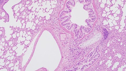
The routine stain in histology is haematoxylin and eosin - H + E. There are many other stains available, which are referred to as special stains. They are used to demonstrate particular features, according to the diagnostic need.
Haematoxylin comes from the heartwood of the logwood tree (Haematoxylin campechianum). Pure haematoxylin is colourless and is readily oxidized to a reddish dye called haematein, which is the active ingredient. Haematein is not entirely stable and gradually turns colourless by further oxidation. In practice, this breakdown is balanced by the oxidation of an equivalent amount of haematoxylin to haematein so that the solution has a useful life of several months to several years. Haematoxylin solutions are only useful after ripening. This will take place naturally after several months of shelf life or can be done artificially by adding an oxidizing agent. Haematoxylin will not directly stain tissues so a mordant (usually aluminium, iron or tungsten) is mixed into the solution. Alum haematoxylin will stain nuclei a dark red colour. This is changed to blue by dipping into a weak alkaline solution and is called ”blue-ing”. Methods of haematoxylin staining are either progressive or regressive. With progressive staining, the slides remain in haematoxylin until the desired amount of staining is reached, before washing and blue-ing. With the regressive staining, the slides are overstained, washed and the excess haematoxylin is differentiated out by dipping in 1% acid alcohol, before washing and blue-ing. The slides are well washed before counterstaining with eosin- failure to do so will result in poor staining and the eosin will be neutralized by the blue-ing solution. Eosin is a synthetic xanthene dye and is usually prepared as a 0.5 – 1% alcoholic or aqueous solution. It is usually made up as a stock solution which renders it less susceptible to moulds and is diluted prior to use. Eosin is an acidic dye with an affinity for cytoplasm. Decalcified tissues and those fixed with mercury based fixatives can sometimes stain very intensely with eosin.
Decalcification
Occasionally bone or other tissue with heavy calcium deposits needs to be processed, sectioned and stained. These specimens require the removal of calcium before sectioning. There are occasions when sections of undecalcified bone are needed ie. studies into metabolic bone disease, but special techniques are required for this.
Removal of calcium (or decalcification) must be done after the tissue has been thoroughly fixed and prior to processing. Large specimens ie. patella, finger etc, will survive the process better, if after preliminary fixation,they are sawn into slices and fixation continued. This enables the inner tissue structures to fix quickly and makes the subsequent decalcification easier. The slices can be trimmed to size for processing if needed.
There are various methods which can be used, none of which are perfect and all will damage the tissue to some degree.
Here at the JCSMR, I use a combination of a commercial preparation called Fastcal, containing hydrochloric acid and a 5% nitric acid solution containing urea. The solutions are not combined but used in conjunction with each other.
The samples are placed in nitric acid in a container with a magnetic stirrer for approximately 2 hours. This is adequate for small bones, ie. mouse ribs or femurs. Larger bones, (rat ankles or femurs), may need several changes of nitric acid. Large specimens eg. mouse heads, should have several hours in nitric acid. Rinse in water and then slice in half or acording to the researcher's wishes, place in labelled cassettes and continue the decalcification.
Tissue must not be left in decalcification fluid overnight, and should be transfered to 70% alcohol. When complete, the tissue is cut to size, placed in cassettes and washed well in running water. The cassettes can be stored in 70% alcohol before being processed on a process cycle commencing with 70% alcohol.
Blocks are carefully trimmed and chilled lightly - too cold makes them brittle and liable to chip. Bony sections are hard to cut and must be floated on to a coated slide (silane, albumin) or they may wash off during staining. Decalcified tissue tends to stain very strongly with eosin and will therefore appear "redder" than the usual soft tissue H+E.
After taking the sections to water, soak the slides in saturted lithium carboate solution for 5 minutes. Wash in running water for 5 minutes before proceeding with the stain of choice. This allows for better staining as it raises the pH of the section.
If tissue is discovered to have calcified deposits/gritty areas when trimming the block, it can be "surface" decalcified. The block is trimmed as much as possible and soaked for a short while in Fastcal before re-trimming. Repeat this step until a full surface of the tissue is reached. It is washed well, dried and cooled lightly. Sufficient calcium should have been removed to enable a few good sections to be cut. They should also be floated on to a coated slide, as a precaution.
Specimen preparation
Do you have specimens for histology? Here's what you've got to do:
1. Place specimen in fixative of choice. Specimen containers are available in all shapes and sizes (70ml capacity) in large bin, under the table in room 1.223.
- Bouins - you make that up yourself, Anne has the recipe if needed.
- Formalin is available in room 1.223.
2. Fill out request form, available in room 1.216.
Details required are:
- Name and contact number.
- Details of specimen type.
- Fixation used.
- Type of embedding required.
- Number of slides required.
- Staining/unstaining and type of stains required.
- Specify if urgent or not.
- Account number for billing purposes.
Specimens must be clearly labeled and left with request form on the trolley in room 1.216. NOTE: For the unusual/exotic specimens eg. bone, paper, plants, insects - please check with Anne first.
Equipment
| Name | Description |
|---|---|
| Tissue processor - room 1.223 |
The tissue is processed on an automatic tissue processor called the Medite Tissue Processing Centre. Normal maintenance includes topping up solution levels when needed, changing solutions regularly to ensure specimen quality and cleaning processing baskets. This instrument is NOT available for use other than by Histology staff. |
| Embedding Centre - room 1.223 |
Tissue is embedded into wax blocks on the embedding centre- a Medite TBS 88 PES - PES stands for Paraffin Embedding System. The instrument requires little to no maintenance other than cleaning up spilt wax and emptying the overflow tray. This instument is not usually available for use other than by Histology staff. However, occasionally slides need to be coated with wax (ie protect unstained sections for transport). If this is needed, just check with histology staff before doing so. |
| Cryostat - room 1.216 |
One Bright cryostat is available for staff/students to use. Booking sheets are provided on each instrument, along with Kleenex, forceps, razorblades and pencils - however please provide your own slides. When booking a timeslot, please leave a phone extension in case you need to be contacted. Knives are provided for the cryostats for those groups not having their own. Knives are sharpened regularly and groups may leave their knives for sharpening as required. The knives are stored in the freezer. A few points to remember:
Safety Notes * NEVER leave a knife in the cryostat after you have finished cutting - always return it to its box. * NEVER leave sharp implements ie scalpel, razor blades or sharp forceps in the cryostat. |
| Microtomes - room 1.216 | There are two microtomes – a PFM microtome which is used ONLY by Histology staff. The other is a Leitz 1512 which is available for anyone to cut their own blocks or to learn on if desired. The hourly charge rate is the same as for use of the cryostats. A booking needs to be made so that the waterbath and ice trays are ready for use. Disposable blades are provided for use, inclusive in the hourly rate. however, you will need to provide your own slides and slide trays/boxes. |
| Waterbath - room1.216 | There is a Labex waterbath capable of heating up to approximately 110° C, used for special stains and for antigen retrieval procedures. Anyone wishing to use the waterbath, particularly for antigen retrieval is welcome to do so, free !!! Please arrange with Anne so that the waterbath is at the required temperature at the time you want (It is not turned on every day, only on an "as needs" basis). |
| Haematoxlin + Eosin (H+E) Stains - room 1.216 | The H+E stains are set up in the fume cupboard. Solutions are changed regularly, depending on the slide numbers being stained. Anyone wishing to de-wax their unstained slides or do an H+E on their own slides is welcome to do so (free, again !). For advice/help on these procedures and help with coverslipping, please see Anne. Please provide your own coverslips. |
| Knife Sharpener - room 1.223 |
The knife sharpener is a Temtool Perma-Sharp and is used to sharpen solid steel blades for either microtome or cryostat. Knives can be left at any time for sharpening. This instrument is NOT available for use other than by Histology staff. |
Techniques
| Name | Description |
|---|---|
|
Cell clot /Cell block preparation |
Urine, blood and fluid aspirations are usually examined by making smears, films or cytospin preparations, before staining and examining microscopically. In the case of fluid aspirations or where fluids have obvious fragments/ cellular deposits, it can be very helpful to prepare a cell clot/ cell block. The two main requirements for cell clot preparation are thrombin and plasma. Thrombin Thrombin is available from pharmacies and is marketed as Thrombostat, made by Park-Davis. It is not usually a stock item and will need to be ordered. Thrombostat is available in two sizes 5ml/5000units and 10ml/10000units, in a vial of powdered thrombin. The 5ml size is most convenient to use. Stock Thrombin Solution Add 5ml sterile normal saline to the vial using a 5ml syringe and needle. Mix gently to ensure powder dissolves. This solution has a concentration of 1000units/ml. Working Thrombin Solution Dilute 1:10 with sterile distilled water. This is easy if 9ml sterile distilled water is added to a sterile tube then 1ml stock Thrombin, cap and mix. This solution has a concentration of 100units/ml. Aliquot into Eppendorf tubes in 500µl volumes, date and freeze. Each aliquot is sufficient for one cell clot. Note: Once the stock Thrombin solution is prepared, it can be stored at 4°C but must be used within the week. If it is not possible to use 5ml stock in the week, freeze the stock solution in ml aliquots. Plasma Plasma should be collected and aliquotted into Eppendorf tubes in 250µl volumes, dated and frozen. Each aliquot is sufficient for one cell clot. |
|
Unfixed or alcohol fixed material |
For alcohol fixed specimens, mix and pour into a labeled centrifuge tube. For fluids, pour 10ml, including any particles or fragments into a labeled centrifuge tube. Spin down for 10 minutes at 1200rpm, decant supernatant leaving pellet undisturbed in the bottom of the tube. Add 250µl thawed, room temperature plasma and gently mix by vortex. If there is only a very small amount of material, mix gently by hand. Add 500µl of thawed, room temperature thrombin and mix gently by vortex. Hold in palm or under arm until set. If doing multiple specimens, place in a rack in a 37°C waterbath until set. Ring clot and ensure it is free from the tube. Add formalin to the tube or tip the clot into a jar containing formalin. The clot is then processed for histology and slides prepared as for tissue. |
|
Formalin fixed material |
Pour tissue fragments, cellular material etc. in formalin into a labeled centrifuge tube. Rinse container with formalin and fill centrifuge tube. Centrifuge for 10min at 1200rpm. Remove supernatant with Pasteur pipette to avoid disturbing sediment. Fill tube with sterile normal saline, cap and mix thoroughly. Centrifuge for 10 minutes at 1200rpm. Remove saline and repeat this step. It is important to remove all traces of formalin or the clot will never set. Remove supernatant, leaving sediment undisturbed and add plasma and thrombin as per procedure for unfixed and alcohol fixed material. References
|
|
Preparation of Heavily blood stained fluids |
Centrifuge in a standard centrifuge tube and discard supernatant. Fill tube with a mixture of 9 parts cytolyt and 1 part glacial acetic acid. Resuspend deposit by gentle mixing and stand for 10minutes. Centrifuge again and process deposit as usual. |
|
Fluids with heavy deposits |
Immediately after cytocentrifuging, spread the circles of cellular deposits with another clean glass slide and place into 95% alcohol/cytolyt or airdry. Fix and stain as usual. |
|
Fluids with scanty material |
If specimen is less than 5ml, make up to 10ml with cytolyt, centrifuge and cytospin as usual. Remember, air dried smears cannot be made from cytolyt fixed material. |
Frozen sections
Frozen sections are required for various reasons, either desirable or obligatory, instead of the routine paraffin sections. They are cut using a cryostat which in layman's terms, is a freezing chamber containing a microtome. A few modifications are necessary for operation at very low temperatures but otherwise it is essentially the same microtome used for paraffin sectioning. Most available cryostats operate at temperatures down to -30°C to -40°C.
Procedures which require frozen sections
- Surgical procedures where a rapid diagnosis is required.
- For demonstration of certain substances which will either dissolve or be destroyed/altered during tissue processing i.e. enzymes, fat, lipids.
- For demonstration of components of the central nervous system to avoid shrinkage.
- For preparation of hard tissue i.e. tendon which are difficult to cut and flatten after processing.
For surgical procedures and enzyme histochemistry, tissue samples must be fresh and unfixed and frozen immediately on collection. This is also desirable for immunofluorescence studies i.e. on skin and renal biopsies but if there is a problem with freezing immediately, the tissue can be stored in Michel's Buffer/ Transport Medium until it is convenient to freeze and cut. This is very useful as it allows the specimen to be collected in one place and sent elsewhere for testing without deterioration.
For fatty specimens, it is advisable to cut off as much fat as possible before freezing the tissue as fat will not freeze until at least -60°C. If excessively fatty tissue must be cut, it is better to prepare imprint slides. This is done by cutting the tissue and, using the freshly cut surface, press it gently but firmly onto a clean glass slide. Do not squash, drag or smear it as the cells will be destroyed/damaged. The slide should be air dried or fixed immediately - depending on the staining procedure, stained and coverslipped. This technique will provide very good cellular detail.
For some procedures, it is desirable, for section quality to fix the tissue first, either in 4% paraformaldehyde or formalin. The fixative is then leached out by soaking the tissue in sucrose - concentrations vary from 7% - 30%, depending on the protocol followed. After soaking for several hours to overnight, blot tissue dry and place in OCT or cryomedium of choice and allow tissue to be infiltrated. I find this easily done by placing tissue and OCT in a 5ml screw top tube and fitting tube onto a rotating mixer overnight. The tissue can then be frozen in fresh OCT and either cut or stored in the freezer.
Freezing the tissue
Tissue can be frozen in several ways, but must be frozen quickly or ice artefact will occur. This makes good sectioning very difficult to impossible. Methods include liquid nitrogen, supercooled freon or isopentane and an acetone - dry ice slurry. The latter is easy and convenient, especially if there are only a few samples and you are not going to be doing very many frozen sections.
Acetone - Dry Ice Slurry
Place a wide mouthed container - stainless steel/ Pyrex which has about 400ml capacity into an esky of dry ice. Pour acetone into the container to a depth of 2cm. Add very small amounts of dry ice to the acetone and TAKE CARE - the acetone will boil furiously until it starts to cool. Keep adding dry ice slowly until a sludge is reached. This will keep for several hours if stored in dry ice. Place molds - either aluminum foil or commercial - onto slurry and add a little OCT. As it turns white, orient your piece of tissue in the OCT. By placing your tissue onto the OCT as it is turning white, the tissue will be held in the position you want it. Fill immediately with OCT so that the block freezes "as one" - you don't want the bottom shearing off when cutting. Keep the mold in the slurry until it is quite set, then store it in the dry ice/ freezer until cutting. Do one block at a time.
When all specimens have been frozen, place the slurry on the bench and it will gradually come to room temperature. The acetone can be used again, but store in a bottle labelled "Acetone for Dry Ice Slurry". It should not be used for any other procedure.
Instructions on how to use and maintain the cryostats and making the slurry are available. Just see Anne in Histology and arrange a time.
For those interested, the recipes for Michel's buffer follows.
Michel's Buffer/ Transport Medium
Solutions required to make Michel's Buffer are:
Stock Solutions:
1M Potassium Citrate
Potassium Citrate H2O 32.4g
Distilled water 100ml
0.1M Magnesium Sulphate
Magnesium Sulphate 7H2O 2.43g
Distilled water 100ml
0.1M N-Ethylmaleimide
N-Ethylmaleimide 1.25g
Distilled water 100ml
1M Potassium Hydroxide
Potassium Hydroxide 5.61g
Distilled water 100ml
Stock Buffer Solution:
1M Potassium Citrate 2.5ml
0.1M Magnesium Sulphate 5.0ml
0.1M N-Ethylmaleinmide 5.0ml
Distilled water 87.5ml
Adjust to pH 7.0 with 1M Potassium Hydroxide
Working Buffer Solution:
100ml Michel's Buffer stock solution
55g Ammonium Sulphate
Dissolve and adjust to pH 7.0 with 1M Potassium Hydroxide.
Store all solutions at 4°C. The working solution will keep almost indefinitely, though it is sensible to make up volumes that are likely to be used within a reasonable time.
For a piece of tissue, approx. 4mm cube, a volume of 2-3ml is sufficient. Whilst solutions should be stored at 4°C, it is not necessary to remain at 4°C whilst being transported. No deterioration will occur to the tissue. On receipt of a specimen in buffer solution, store at 4°C until ready to proceed.
Specimens received in Michel's Buffer need to be washed in a washing buffer, with three changes of 10 minutes each. Place tube on a rotating mixer to ensure all vestiges of Michel's buffer are removed. The specimen is gently blotted before freezing.
Coons Washing Buffer Stock Solution:
Disodium hydrogen orthophosphate 4.25g / 2.125g
Sodium chloride 43.5g / 21.75g
Potassium dihydrogen orthophosphate 2.7g / 1.35g
Distilled water 1000ml / 500ml
Adjust to pH 7.1 - 7.3 with 1N sodium hydroxide
Washing Buffer Working Solution:
Dilute stock solution 1 in 5 with distilled water. Check pH and adjust if necessary.
References:
Michel, B, Milner,Y and David, K.
Journal Invest. Dermatol., Vol 59, 1973, p449, "Preservation of tissue fixed immunofluorescence in skin biopsies of patients with LE and bullous disease"- (Prelim report)
Special stains
Special stains are designated such to distinguish them from the routine stain, H & E. Sometimes they are requested after examining the H & E to demonstrate the presence or lack of a particular feature. Othertimes they form a part of a diagnostic panel and in essence, become routine for that type of tisssue-ie, a liver biopsy will routinely have H & E, PAS I diastase, reticulin, Perls and a trichrome- Massons or Gomori. Some stains will have a control section provided. This is to ensure that not only has the stain been carried out correctly, but also to that the solutions are working. If the control slide is positive, and the test slides are negative, then the feature being tested for is absent.
*Stains where control slides are used are marked with an asterisk.
The range of special stains currently available in Histology are:
| Stain | Use |
|---|---|
| Massons Gomori van Gieson MSB |
Differentiate between muscle and connective tissues |
| Verhoeff Orcein Aldehyde-Fuchsin |
Elastic fibres |
| Verhoeff-van Gieson | Combined stain for elastic fibres, muscle and connective tissue |
| Gordon & Sweet | Reticulin fibres |
| Perls* | Iron |
| Dithizone* | Zinc |
| Rubeanic Acid* | Copper |
| Schmorls * | Melanin |
| Alcian Blue* Alcian Blue-PAS* |
Acid mucins Combined stain to differentiate between acid and other mucins |
| PAS* | Mucins, fungi, keratin, cryptococci and glycogen |
| Alcian Blue* Astra Blue* |
Acid mucins |
| Mucicarmine* | Neutral mucins only |
| Congo Red* | Amyloid |
| Ziehl-Nielson* | Acid fast bacilli, mycobacteria |
| Auramine* | Atypical mycobacteria |
| Gram* | Bacteria |
| Modified Warthin-Starry* Giemsa* |
Helicobacter |
| Warthin-Starry* | Spirochaetes |
| Grocott* | Fungi |
| Modified Methenamine Silver |
Renal basement membrane |
| MGG Giemsa Leishman Diff Quik |
Blood cells |
| MGP* | RNA, DNA, Plasma cells |
| NSE* | Enzyme activitiy in monocytes,histocytes and megakaryocytes |
| Chloroacetate Esterase* | Enzyme activitiy in myeloid cells |
| Modified Aldehyde fuchsin* | Pancreatic beta cells |
| Modified Congo Red Carbolchromotrope |
Eosinophilic granules |
| Modified Giemsa* Toluidine Blue* Astra Blue, low pH* Acian Blue, low pH* Modified Chloroacetate Esterase* |
Mast cells |
| Orcein-Giemsa* | Elastic fibres and mast cells |
| Astra Blue-carbolchromotrope* | Differential stain for mast cells and eosinophils |
| Phloxine-Tartarzine* Amido Black* |
Paneth cell granules |
| Phloxine-Tartrazine* | Inclusion bodies |
| Oil Red O Sudan Black |
Fat, lipids *only on frozen sections |
| Luxol Fast Blue | Myelin |
| Cresyl Violet | Nissl substance, nuclei |
| Luxol Fast Blue- Cresyl Violet |
Combined stain for myelin and nissl substance |
| High Iron Diamine (HID) | Differentiate between sulphonated and carboxylated mucins |
| Melanin Bleach | Used to remove excessive amounts of melanin in tissue, prior to staining with stain of choice |
| Alcian Blue, pH5.8* | Heparan sulphate in beta cells |
There are many other special stains available, though they are not made up as there is or has been no demand for them. if you have a particular feature to be demostrated, come and chek out what is available. Advice and assistance is available for immunostaining on paraffin sedtions. I do not, as a rule, carry out these procedures for staff/students but am happy to help if there are poblems. The waterbath is available at any time for antigen retrieval procedues. If you need to use it, please ring and ask for it to be switched on. It is only turned on an "as needs" basis.
Safety
Histology has a vast number of chemicals, dyes, solvents and solutions, most of which are hazardous to a greater or lesser extent. It can be a dangerous place to work, but with care and commonsense most people survive without any undue problems. (or maybe, they're fixed!)
There are MSDs on file of all chemicals, dyes and solvents in the laboratory. There is also a chemical and stain index which has a record of all chemicals and dyes - with a sheet for each showing an internal reference number, where it is stored and requirements/recautions for handling and disposal.
Some suggestions for safe working in Histology (most are applicable to any laboratory handling dangerous/semi-dangerous goods):
- No eating or drinking in laboratories under any circumstance.
- Wear laboratory gown/coat and gloves when handling chemicals, dyes etc.
- Wear flat heeled, closed shoes-floors in Histology tend to be slippery because of wax. A closed shoe is extra protection from solution spills or falling knives.
- Dispense solvents, chloroform, formalin and acids in fume hoods.
- Never pour chloroform, xylol or any fixative containing mercury (B5, Zenker's, Helley's) down the sink. There are bottles in Histology for all of these. If you have a small volume, you can bring them down and add to our collection. For larger quantities contact the Safety Officer on x53108 about disposal.
- Never pour formalin, formaldehyde, para-formaldehyde or Bouin's fluid down the sink. Place in labelled waste drum in Room 1.223.
- Bouin's solution is made with Picric Acid and formaldehyde. Picric Acid must never be allowed to dry out as it is explosive when dry. When making up Bouin's,
always ensure there is plenty of water to cover the crystals before sealing tightly. - Mop up any spillage immediately.
- Formalin Spills - small amounts can be quickly mopped up using paper towel, towels etc. These should be rinsed in the fume hood to eliminate the smell.
For larger spills, sprinkle Polyform F granules around the edge of the spill and then cover the rest of the spill. Polyform F will absorb the fluid and fumes and should be left for at least half an hour before cleaning up into a waste bag. The floor will be very slippery afterwards and must be scrubbed with hot, soapy water. Polyform F is provided in the Processing Room, next to the formalin container and in the main laboratory, in the fume hood. - Always turn off the formalin tap when you fill your bottles. A slow drip can leave a big spill, especially if it is overnight.
- Be aware of and practice OH&S requirements for your own and fellow workers' health and wellbeing.
Handy hints
Tissue colour markers
It is easy, when examining an intact specimen, to identifiy specific areas, margins, excision lines etc. However it is not always so obvious microscopically unless it has been identified or marked. This is easily done by painting the tissue with a dye which is impervious to solvents and will provide a contrast to the stain to be performed. It can also be used to identify tissues from several animals/specimens in the one block. Dyes suitable for use are: Indian Ink, Mercurochrome, Alcian Blue, Alcian Green and Calli Calligraphy ink- black or green.
Coverslipping Hints
The most common problems in coverslipping are airbubbles and too little mountant. This becomes evident some time after coverslipping and results in large airspaces under the coverslip.From experience, I find it is easier to transfer the mountant to a small dispenser bottle - a sauce bottle or a clean dried OCT bottle is ideal. Dispensing the mountant is easy, there is control over the volume used and it is not so messy. When not in use, the bottle cap must be kept on or the mountant will thicken up. An ideal amount of mountant is a line about 1/2 to 2/3 the length of the coverslip.If the slide is airdried - Dip the slide in xylol and gently drop onto a prepared coverslip. Wait a few moments before gently tapping out the bubbles. If the slides are in xylol - Prepare coverslip, drain slide briefly and drop on to coverslip. Wait and then tap out the bubbles. The drier the mountant is on the slide, the harder it will be to spread and move the bubbles. If it is too dry, throw out the cover slip and start again. Do not allow slides to dry too much after removing from xylol - you tend to use too much mountant in order to make it spread evenly under the coverslip. If the mountant in the OCT bottle becomes thickened and hard to dispense, add a little xylol and beat in. Keep adding xylol and beating until the consistency is right. As the mountant will be full of fine air bubbles, let it stand overnight and these will disappear. Aqueous mountants are not as easy to use as they do not have the viscosity of the solvent based mountants and consequently, the air bubbles are very difficult to move. You need to be more generous with the mountant and try not to shake the bottle too much. Too much movement creates bubbles. Make sure there is plenty of water on the slide before dropping on the coverslip and try to ensure any bubbles are pushed away from sections/smears etc.
Do you have old slides that need recoverslipping and/or restaining?
Soak slides in Xylol (or water if applicable) until the old coverslip floats off and the original mountant is dissolved. The time taken for this depends on how long the slides have been coverslipped and may require gentle heat ( not warmer than 40° C) and several changes of solution. If you need to warm the solution to speed up the process, make sure the container is well sealed. Hot xylol fumes are not pleasant. When slides are ready, recoverslip. If the stain is faded, it can be bleached out (and NOT by using domestic bleach!) and either the original stain re-applied or another stain done instead. This only applies to H & E or special stains - not immunostains.
Histology Library
There are a range of books in Histology covering all topics in basic histology, special stains, dyes, histochemistry, immuno staining and two histology atlases. These atlases are very useful, with coloured plates of normal and some abnormal tissues. These books are available for consultation and can be borrowed at any time for at least two weeks with longer time if needed. Please feel free to come and check them out. The most frequently borrowed books are the atlases.
|
Histology |
Internal (JCSMR) |
External (within ANU) |
Outside ANU |
|---|---|---|---|
|
Block-processing only |
$5 each |
$5 each |
$7 each |
|
Slide, cut and stained |
$5 each |
$5 each |
$7 each |
|
Slide, cut and stained, multiple blocks or levels per slide Slide, unstained, multiple blocks or levels per slide |
$7.50 each
$5 each |
$7.50 each
$5 each |
$10 each
$10 each |
|
Slide, unstained |
$2.50 each |
$2.50 each |
$7 each |
|
Slide, staining only |
$2.50 each |
$2.50 each |
$7 each |
|
Cryostat knife re-sharpening |
$50 each |
$50 each |
$60 each |
|
In situ blocks, processing only |
$10 each |
$10 each |
$12 each |
|
In situ slides |
$5 each |
$5 each |
$7 each |
|
Cryostat/microtome |
$7.50 per hour |
$7.50 per hour |
$15 per hour |


